
Case Courtesy Dr. Peter Kim Nelson
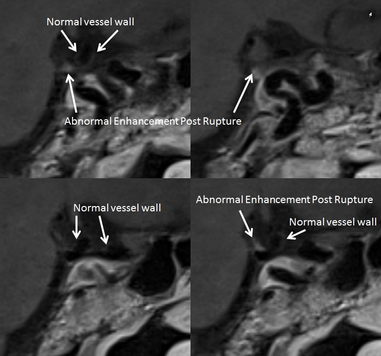
MR vessel wall imaging is far from mature. What role it will ultimately play is still unclear. However, one consistent thing it seems to capable of is pointing to the ruptured aneurysm, when more than one is present. There is usually more enhancement around the rupture site. What tissues are enhancing is not too clear, since aneurysm wall must be focally absent by definition. Is it thickened wall adjacent to rupture site? Tissue “inflammation” around hematoma? How soon does it develop and how long it persists? Lots of questions. But there is potential
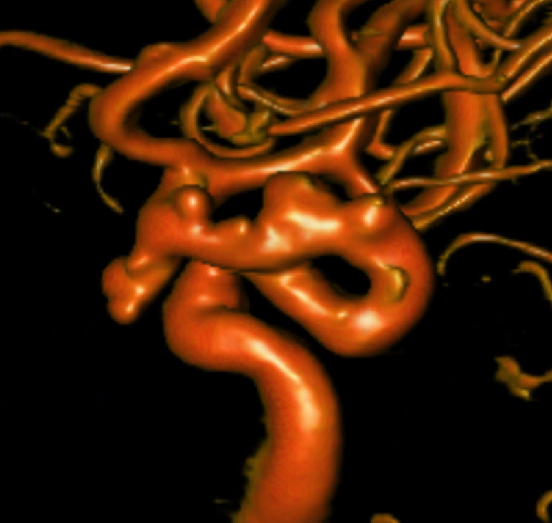
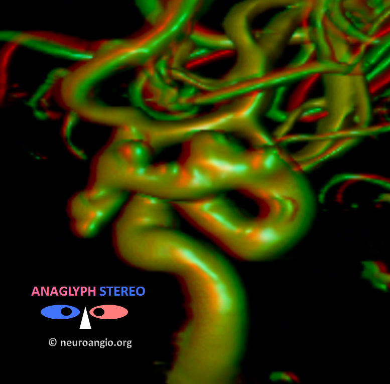
Patient presents with subarachnoid hemorrhage several weeks prior. Angio shows “status aneurysmaticus” — a picture perfect poster for the idea that aneurysms are consequences of underlying segmental deficiency. The entire segment is diseased, with a range of aneurysm sizes, locations and shapes.
Statistically, the PCOM is by far the most likely suspect — it is the largest, morphologically most irregular, and is a PCOM . Vessel wall imaging agrees
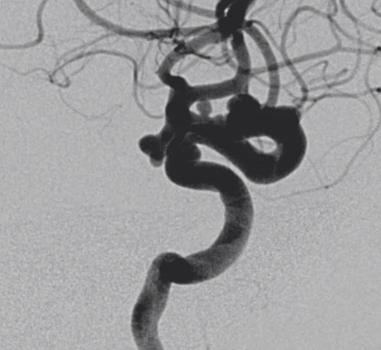
Angio
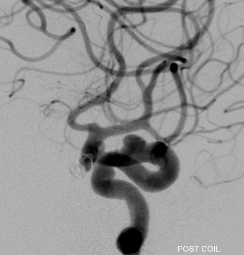
A few coils are placed into the sac to secure the dome
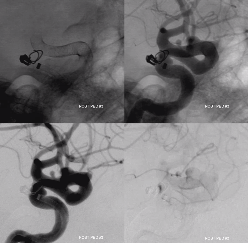
Followed by Pipelines to treat the whole diseased segment
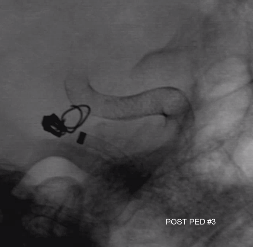
Post
Some literature
https://www.ncbi.nlm.nih.gov/pubmed/29902602
https://jnis.bmj.com/content/early/2019/07/23/neurintsurg-2019-014938