
How to deal with a femoral puncture site pseudoaneurysm? First line is ultrasound-guided compression. It is straightforward and highly effective.
- Take a curved abdominal probe
- Put it over puncture site and turn on color
- Wiggle probe around until you can see the tract from femoral artery to the pseudoaneurysm
- Push down on probe until Doppler flow in pseudoaneurysm and tract disappears, while that in femoral artery persists
- Hold steady for 15-20 min and don’t peek to see if its working. It hurts the patient a lot. Consider giving pain meds in advance. The good news is that your fingers will hurt also.
- Let go slowly and check to see if tract and pseudoaneurysm flow are gone.
- If not, consider repeating above 1-2 more times before doing something else.
- If success, leave patient in bed with leg straight for a while
- Next time, think radial
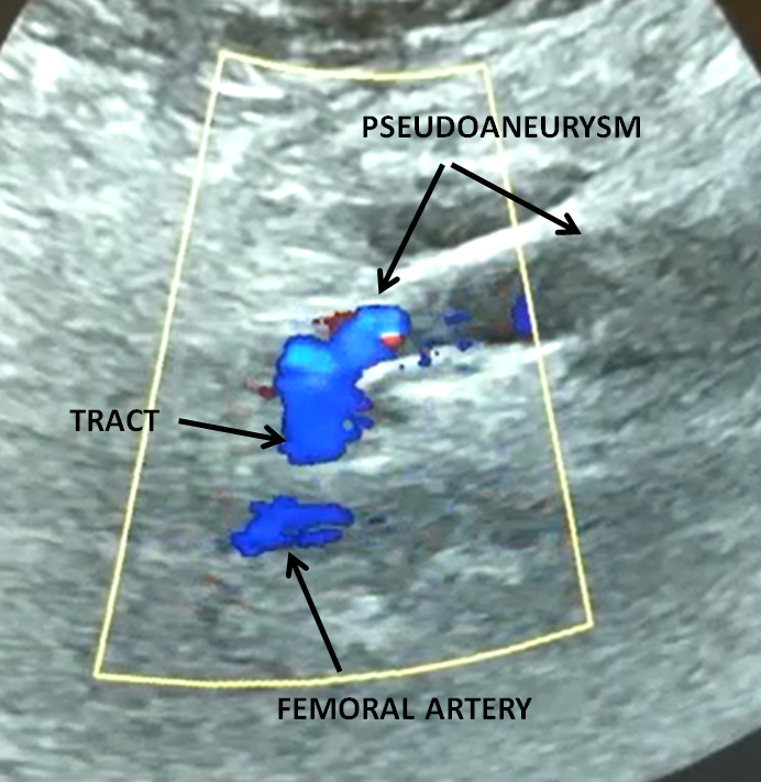
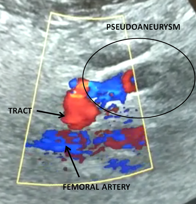
Here is a typical ultrasound picture of pseudoaneurysm
Here is a video of how to treat it
Use it in your stubborn femoral access practice, or see list of radial cases here